Monthly Archives: April 2011

ADEPT – A way to target drugs to tumours with minimal collateral damage.

Hello there.

The focus on this post will be on a method that should facilitate the accurate delivery of drugs to tumours, this is important because quite a few cancer chemotherapeutic drugs, which are lethal to cancer cells, are also lethal against normal cells and can have adverse side effects. This is a problem because the toxicity of drugs to the patient limits how much can be given.

The key question to be asked when dealing with this issue is if one can administer the drug just to cancer cells without exposing normal cells to it. ADEPT, which stands for Antibody Directed Enzyme Prodrug Therapy, is one possible way this can be done provided the cancer being treated is amenable to this kind of treatment.

One of the basic concepts involved is that cancer cells may express antigens or proteins in high levels that are only expressed at low levels in cancer cells. This means that antibodies that are targeted against these antibodies will find themselves preferentially binding to cancer cells, stick something to the antibody and the payload will be delivered only to cancer cells. This is important for ADEPT but by itself is not enough.

Antibodies that are bound to something are called immunoconjugates, while sticking toxic drugs to antibody molecules may increase the efficacy of targeting, these drugs may still exert side effects on account of having to be circulated through the body. This leads one to look for ways to activate the drug only in the vicinity of the tumour, and ADEPT does this.

Antibodies that are used in ADEPT are chemically modified to accommodate a catalytic subunit from an enzyme (enzymes are usually protein molecules that increase or decrease the rate of a chemical reaction, one example would be papain from papayas which is used in tenderizing meat). There are some enzymes which can be used to convert a non-toxic, inert form of the drug to an activated, toxic form through chemical reactions, and this forms the second of three components required for ADEPT.

The non-toxic precursor molecule that is later enzymatically activated is called the Prodrug. The antibody, the attached enzyme and the prodrug form the troika that is required for ADEPT to be employed successfully.


Protocols for ADEPT, an outline.

In a typical ADEPT procedure, the following happens.

[1] Antibodies specific to an antigen overexpressed by antibodies may be produced using Monoclonal antibody technology (there is a blog article I’ve written on that technology on this blog, you may want to go through it)

[2] A suitable enzyme is selected for the conversion of a prodrug to an active drug, this is then chemically engineered to fuse it with the antibodies derived during step [1]

[3] The enzyme-Mab conjugates are then injected into the body where they find themselves concentrating at tumours.

[4] The prodrug is then injected and circulates throughout the body, because this is inactive and inert it won’t cause side effects during this stage.

[5] When the prodrug encounters tumours, which have a high concentration of activating enzymes due to antibody-directed binding, it is converted to the active form which is capable of killing tumour cells. This means that it is possible to have drugs exerting their effects only at the location of tumours, thus preventing problems with toxicity during systemic circulation.

This may be further improved by using choices of enzyme-prodrug pairs where the enzyme only efficiently converts the prodrug to the active drug in the microenvironment of the tumour, which can be different from the environment in normal tissues. One example of such varying conditions is the presence of Hypoxia (low oxygen supply) in tumours.

How ADEPT Works (A diagram I made) - Antibodies, which bind preferentially to cancer antigens, have enzymes fused to them, which activates a non-toxic prodrug to an active drug only where tumours are present, increasing efficacy and lowering toxicity to normal cells.

Please click on the thumbnail for a larger image. I know it is a crap illustration but I promise to produce better ones if someone buys me a graphics tablet :P.

This is a very short post since the strategy is clinically very nascent, you may however find this list of literature useful in learning more about the topic.

Potential Problems with ADEPT.

[1] The drug may cause toxicity after activation in tumours until it is excreted.
[2] Evolution of drug resistance, chemo-resistant cancer cells are able to express genes like Mdr1 which can pump drugs out of cells, this means that ADEPT will be useless if the ultimate activated product is ineffective.
[3] Evolution of cancer cells that have downregulated expression of the antigens that bind to the antibodies being used, which is going to prevent adequate concentrations of the enzyme from building up, and consequently the activated drug from being in effective concentrations, in the vicinity of the tumour.

Other approaches to Enzyme Prodrug Therapy.

One may use hormones conjugated to the enzyme to target cancer cells that overexpress a particular hormone receptor, such as androgen receptor 2 in prostrate cancer cells (this is hypothetical), alternatively, genetic engineering could potentially be used to make only cancer cells express the enzyme that then converts the prodrug to the toxic, active form, thus killing cells that have been genetically altered. This is called GDEPT (Gene Directed Enzyme Prodrug Therapy)
and I may elaborate upon this in a future post.

It is also possible to use conditionally replicative viruses to deliver GDEPT to cells, but this variant, since a virus is used, is called VDEPT (Virus Directed Enzyme Prodrug Therapy) You may find the following paper a good read.

” Strategies for Enzyme/Prodrug Cancer Therapy, Xu and McLeod, Clinical Cancer Research, November 2001 7; 3314 ” which you may access here

You may also find this paper which details a clinical trial featuring the use of ADEPT to treat Colorectal Carcinoma, titled
“A phase I trial of antibody directed enzyme prodrug therapy (ADEPT) in patients with advanced colorectal carcinoma or other CEA producing tumours” a useful read in understanding a case study in ADEPT, which introduces you to a candidate prodrug, a candidate enzyme and a candidate antibody, examples of which I personally did not provide in my post.

Finally, here is the page of a lab at University College London’s Cancer Institute that specializes in this area.

I hope you found the article a good read. That is all from me until the next post.

-Ankur.

Science Primer – Winning the War on Malaria.

Malaria is a disease that is caused by protozoans of the genus Plasmodium. The most common species seen causing this disease are Plasmodium falciparum and Plasmodium vivax.. If you look at the WHO Executive Summary on the subject of Malaria, which you may duly find here you will see that there were around 225 million cases worldwide reported in 2009 with 900,000 odd deaths from it in 2009.

Now these numbers are staggering and indicate a severe healthcare problem. Current approaches involve the use of insecticide treated mosquito nets and drugs like quinine and combinations of artemisinin with other drugs, but drug resistance is a problem with malaria. It would make great sense to look for ways that subvert the ability of Plasmodium spp to evade drugs and also to find low-intervention, high-impact ways of dealing with the transmission, which occurs through mosquitoes.

In this primer, I would like to introduce you to two landmark studies which offer strategies that may prove immensely useful and effective in the war on malaria.

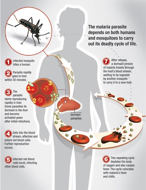
Malaria Life Cycle - An explanation for laymen. Courtesy Metrohealth.org.


[1] Inhibiting PAK-MEK Signalling in Erythrocytes Kills the Parasite.

Now Plasmodium spp (species) are intracellular (inside cells) parasites, and this often implies that there will be molecular interactions between the cells of the host and the cells of the parasite, and in some cases this opens up targets for therapy on the host side of things.

I wish to introduce a paper here, which should make for illuminating reading, titled “Activation of a PAK-MEK signalling pathway in malaria parasite-infected erythrocytes, Sicard et al, Cellular Microbiology, doi:10.1111/j.1462-5822.2011.01582.x” which you may duly access here

The authors of the work report certain things that are of importance in the fight against malaria. They report that there is a group of three enzymes that comprises the core of the signalling pathway they investigated, MAPK (aka ERK) , MAPKK (aka MEK) and MAPKKK (aka MEKK), all upstream of MAPK/ERK.

That is, if you took the core module, it would work like this

MEKK activates MEK activates MAPK.

MEK can also be activated in the absence of MEKK, by an enzyme called PAK, which gives us two potential links in the chain to knock out. The particularly interesting thing to come out of the study is that they managed to identify that these pathways are stimulated by P.falciparum infection of erythrocytes (or RBCs) and that there are no clear orthologues (malarial genes that do the same thing as the human genes mentioned above) in the P.falciparum genome. They also tested if this attribute was evolutionarily conserved in other members of the genus by analysing the genome of a species of Plasmodium that infects mice. This was done to see if P.falciparum inherited orthologues from its ancestors and then lost it or if the genus never had it in the first place. They found that the latter was true.

So we now have a target in the cell that malarial parasites need to use but don’t have a usable version of those targets themselves, it is like a person who cannot breathe needing a ventilator, take away the ventilator and the person is a goner.
In this study, the authors investigated if they could take away the signalling pathway safely to kill the parasite.

They treated cell cultures that were infected with P.falciparum with drugs that block the signalling pathway, and found that this halts the growth and development of the parasite in RBCs, and since this is a critical step in the life cycle of the parasite it cannot develop further, thus stopping the infection in its tracks. Inhibitors that work to block these pathways allosterically (that is, by modifying kinases upstream in the signalling pathway) were found to kill these parasites while being in concentrations that didn’t harm cells.

There are several drugs in development and use for cancer that work by inhibiting the same signalling pathways, the study suggests that a repurposing of those drugs to treat malaria could be a very effective strategy. They also found that it can disrupt other stages of development of the malarial parasite, both in the case of P.falciparum and in the case of the rodent-infecting-species (P.berghei)

I hope you read the paper to know more about the hows, whys and whats of the study. I will now move on to the next part of the article.

[2] Transgenic mosquitoes that are resistant to malaria can be engineered to take over populations.

Current approaches that involve a focus on killing mosquitoes (especially Anopheles spp) aim to work by preventing transmission of the parasite from human to mosquito and vice versa, thus stopping it from spreading in the population and infecting people.

I now wish to introduce you to advances that take this approach further with great effect.

Now originally the problem was that if you inserted a copy of a gene that imparted resistance to malaria in male mosquitoes, it would be difficult to expect it to take over the population unless there was a strong selective pressure acting on those mosquitoes. To get round this, scientists have developed something called a homing endonuclease that carries a modified mosquito gene. The endonuclease cuts the DNA of the mosquito at a certain location and the mosquito, when it repairs the break, will use the modified version of the gene that has been put in to patch it up, and this modified gene then gets into all sperm of modified mosquitoes instead of about 50% with the traditional approach.

They carried out a caged study on an experimental population and found that genetic modification was easily able to spread through it, which opens the door to any approach that may seek to modify mosquito populations such that they are not amenable to spreading malaria using small amounts of transgenic mosquitoes to introduce desired traits into the population.

For a popular science account of this, please see the BBC News article here. For a detailed scientific treatment of the homing endonuclease research in question, please see this paper from Nature (which unfortunately is stuck behind a paywall)

I have however been able to locate a copy on the web that is available for access from the University of Washington, you may duly find that paper here
which looks like a cracking read (don’t take my word for it, read it yourself :P)

So we now have potential methods that can kill the parasite in case of infection and can prevent infection in the first place by using genetic engineering in combination with knowledge of inheritance. We could be very much on the verge of a breakthrough, science could soon be saving millions of lives. I also wish to make a mention of the very cool technology that enables malaria to be diagnosed with the help of a cellphone at this juncture, which you may read about here

Cellscope - The device that may change the way diseases are diagnosed in rural areas.

That is all from me on this science primer, see you soon with one more.

– Ankur “Exploreable” Chakravarthy

Intolerant atheists never happy.

Hello 😀

So this morning I was searching the news and found a fun little article on the Telegraph website, entitled bodly:

‘THE INTOLERANT ATHEISTS WILL NEVER BE HAPPY’

After a quick read I have determined that a more appropriate title would be:

‘THE IRRATIONAL CHRISTIANS NEVER PRESENT A FAIR ANALYSIS OF A SITUATION, AND INSTEAD JUMP TO ACCUSATIONS OF RELIGIOUS DISCRIMINATION’

Or there should at least be a little disclaimer in there. But of course, the writer of this ridiculous anti-atheist tirade is entitled to express her opinion, and in response, I’m going to express why she’s wrong.

So she opens her article by explaining that:

‘Celebrating a holiday in Britain is like trying to celebrate it in an unhappy family. The best-laid plans for reviving much-loved traditions quickly blow up in an almighty row. There’s no embarrassing uncle in his cups or stroppy in-laws; just schools that drop Nativity plays, shopping centres that phase out carols, and offices that shun Christmas trees. When Christians meekly complain that their Christmas is being ruined, the powers-that-be shout them down: “It’s in the name of diversity, stupid!”’

..whilst not actually pointing to any examples of where this has happened. I personally know of no atheists that would like to see Christmas disappear, and whilst for many it has lost its religious significance, indeed it is a tradition that many of us have grown up with, that is very close to our hearts; not least because it gives us all time off work, is an excuse to get PRESENTS, and is one of the rare times of year that families can all get together and let their hair down. Having said all that, if someone wants to protest Christmas or other religious holidays, then they are entitled to do so, and I honestly don’t care if it upsets people. After all, it seems that what this woman is saying is that atheists should shut up and submit to what Christians say for no good reason. Surely there’s a double standard going on here? Sorry Cristina, but your rights end where others’ begin. I’m all for religious freedom, as this is what allows me to express my atheism. I for one would hate to live in a country where people risk hefty fines for ‘crimes’ of blasphemy, and face being cast out by their families or even being murdered for simply not accepting the existence of a deity.

But to the main point of the article:

‘Now there’s a new rumpus, and just in time for Easter. The Wakefield and District Housing Association in West Yorkshire has ordered one of its electricians to remove a palm cross from the dashboard of his company van. *Colin Atkinson, a grandfather and former soldier, faces the sack for refusing to follow orders.’
*not too sure why that part is relevant to her article…

The first thing to point out is that on the large scale of things, this really has absolutely nothing to do with Easter; she is just using this case as an excuse to atheist-bash, when there is actually no evidence to support her claim that the company boss (pictured right) is an atheist at all. Secondly, company policy prohibits employees from displaying personal items in the company’s vehicles. Thirdly, the company is completely within its rights and within the law to demand that he removes the cross, and if he doesn’t he should absolutely face disciplinary action. This is called consistency.

The law states that:
Discrimination on the grounds of religion or belief is prohibited by the Equality Act 2010. Under the Act, it is unlawful for an employer to:

(a) directly discriminate against an employee by treating him or her less favourably than it would treat others because of religion or belief; and

(b) indirectly discriminate against an employee by applying a policy/provision, criterion or practice that disadvantages employees of a particular religion (unless the employer can objectively justify that policy).

The company is quite clearly doing neither of these things, and I suspect that if another employee were to display a symbol representing another religion, they would be asked to remove it as well. The reason for this is quite simple. The cross may be interpreted as representing company values, and the company would rather not be associated with any particular religion because it causes controversies like this one! Furthermore, the company is a housing association, and I’m pretty sure they’re targeting their services at everyone, not just Christians, and they risk alienating people of other faiths if their employees are allowed to display crosses in their vans.

The writer goes on to state that:

‘Clearly, in the eyes of this publicly funded body, Mr Atkinson’s palm cross is on a par with a swastika, or a racist slogan. The symbol of Christ’s ultimate sacrifice strikes Mr Atkinson’s bosses as offensive: any show of Christian allegiance could drive a divisive wedge into this multicultural society.’

Which is literally the most stupid thing I’ve read today, for the reasons previously stated. Ms. Odone is clearly in dire need of a lesson in Logic one-oh-one. They’re not ‘offended’, it’s just against company policy. Simple as that. Making reference to ‘Christ’s ultimate sacrifice’ just shows what pathetic propaganda her article is. We all know what Christians believe, and sadly for them (I can’t believe I’m saying this again) it is completely irrelevant to 1) the law, and 2) the company’s policy.

Now we reach the slippery slope conclusion of the article:

‘At stake is not just a happy holiday. Once banning Christian symbols becomes accepted practice, the rejection of Christian beliefs is next. Already, social services have stopped a Christian couple from fostering children lest they infect their charges with an anti-gay attitude. Soon, the authorities will forbid conscientious objection: Christian doctors, for instance, will be forced to carry out elective abortions, which they regard as a sin.Where will it end? I fear intolerant atheists will not be satisfied until they’ve driven faith underground: Christians, Jews and Muslims will be forced to resort to Masonic handshakes and hush-hush gatherings. Meet you in the catacombs.’

Seriously? This stuff is laughable. For a start, no one is banning religious symbols, and nope, Christian beliefs haven’t been rejected across the board either. Although she has yet to make the case as to why they shouldn’t be rejected. They are extraneous to modern society, even stunting it; causing people to (as she points out) have archaic anti-gay attitudes, misogynistic attitudes, and anti-progress attitudes (to name but a few); warping people’s minds to the extent that, like this woman, they think it’s OK to have homophobic people fostering vulnerable kids. I’ve written an article on this particular case before, which unfortunately was lost with my last blog, but as I pointed out then, what would happen if one of their foster-children turned out to be gay? I suspect they’d be tossed straight back into the orphanage and the so-called caring foster parents wouldn’t look back.

Cristina Odone is apparently ‘a journalist, novelist and broadcaster specialising in the relationship between society, families and faith.’ It seems that once again, religion has produced someone who is incapable of producing a logical argument, or seeing the perfectly good reasons behind a decision if it comes into conflict with her faith. I guess we shouldn’t be surprised.

Now surely if her writing is getting published, mine should?! What do you guys think?!

Juliet 🙂

An introduction to the Cell Cycle.

The Cell Theory, initially proposed by Schleiden and Schwann, puts forth one of the fundamental tenets of biology, which is that all life is cellular, and that these cells propagate their lineages through the process of cell division. Now while there is a process called meiosis that is involved in sexually reproducing organisms, and this is a different form of cell division, we may safely ignore it for purposes of this blog post, which is on the Eukaryotic Cell Cycle.

The Cell Cycle describes the life and times of your typical cell. It consists of several phases which undergraduate students are usually required to acquaint themselves with.

Cell Cycle Summary

Phases of the Cell Cycle.

Once a cell takes birth from its parent cells by division, it enters a Gap phase, in case the cell is committed not to divide further it enters a resting state, marked by G0 , if it does have a fate that entails division however, it stays in the G1 phase, where it tends to perform its normal cellular role and also undergoes cellular growth, including production of organelles and duplication of materials with the exception of chromosomes.

This is then followed by the S phase, where the DNA in a cell is duplicated, this is then followed by the G2 phase where the duplicated DNA is checked for errors and suitably repaired by DNA repair pathways. This is then followed by the Mitotic or the M phase where actual cell division takes place, the daughter cell then enters the G0 or G1 phase and the cycle continues and so on and so forth. G0,G1, S and G2 can all be collectively called the Interphase.

The M Phase (Mitosis)

Now this is something that gets repeated and repeated ad nauseam through the course of biological education. I wish to reiterate the process one more time. 😛

The M phase itself is divided into four phases on the basis of definite cytological characters that may be observed during the process.

Prophase.

In this phase the nucleus disappears, chromatin begins to condense into chromosomes and spindle fibres begin to appear.

Metaphase.

Sister chromatids, joined at the centromere, line up on the Metaphase plate.

Anaphase.

Sister chromatids separate and one of each moves towards the poles, driven by the contraction of spindle fibres.

Telophase

The Nucleus reappears, the mitotic spindle apparatus disappears and a cleavage furrow begins to form. This is more or less the reverse of Prophase. This is followed by the division of the cytoplasm to form two daughter cells.

The division of the nucleus is called Karyokinesis and the division of the cytoplasm is called Cytokinesis.

Fluorescent visualization of the key stages of mitosis, courtesy Molecular Biology of the Cell, Alberts et al.

You can see a video of mitosis occurring, visualized by fluorescent microscopy, below.

How is Mitosis Regulated.

The regulation of mitosis happens through the interaction of proteins called Cyclins and Cyclin Dependent Kinases which are only active in the presence of Cyclins. These proteins act in concert at specific points in the cell cycle, called Cell Cycle Checkpoints.

This will however be described in a future blog post. This concludes a very short (by my usual standards) post for laymen introducing them to fundamentals of basic cytology. You may want to read about topics such as Cell Cycle Checkpoints, how Cyclins operate and how the process is regulated by looking around on the web until I do come up with a post on this topic.

Dividing Chromosomes, courtesy Welcome Images

Bean Root Squash showing Mitosis, courtesy Wellcome Images

Human Cells in various stages of mitosis, fluorescent microscopy, courtesy Wellcome Images

That is all from me as far as this post is concerned.

– Ankur.

Paper Review – A census of amplified and overexpressed human cancer genes

Right, it is not often that I write two paper reviews in a row but I think this paper is interesting enough to review. The paper in question is “A census of amplified and overexpressed human cancer genes, Santarius et al, Nature Reviews Cancer, doi:10.1038/nrc2771”. I will be back to writing non-specialist articles for laymen from the next article I post.

Now unfortunately the paper needs institutional access (NRC and its paywalls) so you will have to find a way of getting a copy of the paper yourself. The apposite NRC page where further access may be sought is here

The abstract provides a decent introduction to what is covered in the paper, I will elaborate upon the details later in the post. I quote the abstract verbatim.

Abstract | Integrated genome-wide screens of DNA copy number and gene expression in human cancers have accelerated the rate of discovery of amplified and overexpressed genes.

However, the biological importance of most of the genes identified in such studies remains unclear. In this Analysis, we propose a weight-of-evidence based classification system for identifying individual genes in amplified regions that are selected for during tumour development.

In a census of the published literature we have identified 77 genes for which there is good evidence of involvement in the development of human cancer.

Introduction

This paper describes a system that outlines criteria for using evidence reported in the literature to identify genes within amplified regions that are selected for during cancer (the fact that something is selected for in cancer cells is a strong pointer to it being critical to disease phenotypes)

The problem that the authors were addressing was that while there are regions that are significantly amplified in cancer cells (that is, the number of copies of particular genes, called the copy number is increased relative to normal cells by means of duplication of chromosomal regions), it is difficult to identify which genes in these amplified segments (called amplicons) actually drive cancer. They note that until this paper was published, the Cancer Genome Atlas had just a paltry six entries for genes that were causally implicated in cancer due to amplification and consequent overexpression. This was as opposed to the sum total of three-hundred-eighty-four genes that were causally implicated in cancer through other mutagenic processes. They emphasize that this shortness of the list was due to lack of reliable data.

In the context of the paper, they have defined an amplified gene as that which has a somatically acquired increased copy number and is overexpressed because of that. They note that there may be varying levels of evidence to establish this link, in some cases we may have data associated just with genes being in an amplified region, which tells us nothing, in some cases we may have evidence showing that amplification is linked to overexpression, in some other cases we may have stringent data showing that knocking such genes out perturbs a cancer phenotype, or that copy number is correlated with clinical outcome. It makes sense to try and put all of this into perspective by developing a system that takes the strength of evidence into consideration, and such a system is what the paper describes.

Details of the Classification System.

The system assigns points to genes for which evidence is present. I present a graphical summary of the scoring and classification system below, I think it does a good job of making details of the classification system clear.

Summary of the classification & scoring system, courtesy Santarius et al (DOI mentioned earlier in the article)

I think the table is pretty much self-explanatory; it is however useful in my opinion to reiterate that in this system all types of evidence are awarded one point and are weighted equally. Class IV genes have just evidence of being in an amplified region, Class III genes have to score at least 1 point, Class II genes 3 points and Class I genes must be demonstrated, preferably in clinical trials, to be viable therapeutic targets where blocking the gene or the gene product improves clinical outcome.

Class I genes can therefore be said to be integral to pathogenesis by amplification, Class II genes could also potentially be implicated and Class III genes require further study. Now I will go back to talking about heterogeneity, the authors have pointed to instances in the paper where different genes within the same amplicon (amplified region) act as drivers in different types of cancer, for example, they point out that in Lobular Breast Carcinoma, the gene FGFR1 is a driver while conventional wisdom deemed two other genes in the same amplified region as better candidates. This has led the authors to classify genes using this system on a per-cancer basis.

Results & Comments

The analysis only includes amplified unmutated (normal) genes & does not include genes that are both mutated and amplified, nor does it include miRNA (microRNA) which may also be involved in dysfunctional expression and can be treated as normal genes that are amplified. For a brief introduction to miRNA please read my blog post on RNA Interference on this very blog.

The authors have documented 62 Class III genes, 12 Class II genes and 3 Class I genes. I fully expect the number of genes in these classes to go up as more and more data from integrated genomic studies start to flood in. The system may prove particularly useful in guiding research programmes by informing further research and what evidence needs to be investigated. It may also enable the identification of overexpressed genes that are integral to cancer progression and thus offer a way of identifying drug targets.

I will now go on to post a table and a diagram of the results the authors have presented in the paper. You may bring up larger versions of these images by clicking on them.

Table of Results by Class & Cancer Type, courtesy Santarius et al.

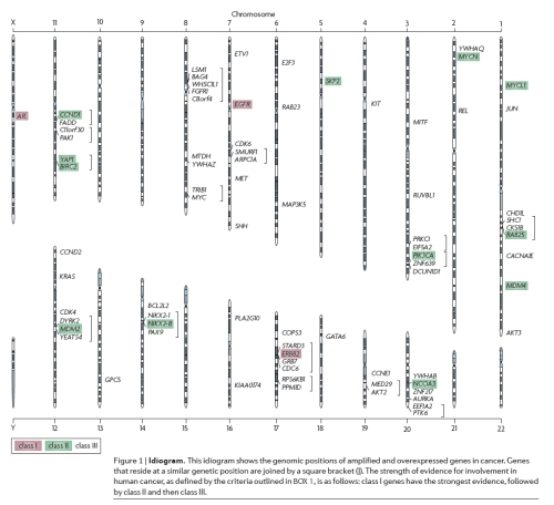
The whole lot can be visualized using a virtual karyogram/idiogram, which follows below.

Graphical Summary of the Chromosomal Locations of the genes implicated in the progression of cancer during the analysis.

Further data about what evidence was used in the classification and scoring of amplified genes may be obtained from the supplements. You may find these supplementary materials here

To conclude, I think this will show you how oncologists may try to make sense of information stemming from a wide range of studies that all provide evidence to different extents and of different types. I also hope that you will be able to visualize how such systems can help organize, compare and utilize data to guide things that range from what studies one may wish to carry out to what targets drug development programmes may look at in the quest for the development of safe, effective targeted therapies against cancer.

That is all from me as far as this paper review is concerned, I hope you enjoyed the post and the paper, and I also hope that you will take time to try and know more about the Cancer Genome Project and how it is helping us understand cancer in all its complexity.

– Ankur.

Paper Review – Selective cell death mediated by small conditional RNAs

Alright then. This is the second paper review I’m writing for the blog. This details a novel method of killing cancer cells selectively. Here, only cancer cells that express a particular gene aberrantly will be killed off through the activation of an innate immune response. This paper details an in vitro study carried out using cell cultures.

You may find it useful to have a copy of the paper handy, and you may find the PDF available from PNAS here

In this paper, the authors elaborate upon a method that uses the physical properties of RNA molecules to drive a chain reaction involving generation of an active molecule of RNA which then goes on to kill cancer cells by apoptosis through the activation of the PKR pathway.


Please note that you will be able to bring up bigger versions of the images you see by clicking on them.

The Actors of the Piece.

PKR (or Protein Kinase R) is a protein that is involved in an immune response to the presence of viral double stranded RNA (dsRNA), it triggers inhibition of protein synthesis and apoptosis when it dimerizes (two molecules of this combine to form one unit) in association with double stranded RNA that is longer than 30 base pairs.

Small Conditional RNA (or scRNA) – These molecules are designed to stay inactive in which a cancer-specific marker is absent, however, when such a marker is present (this shall be referred to as the cognate marker from now on) they will react to form a long double stranded RNA sequence which will go on to activate PKR mediated apoptosis.

The molecules they used in the paper have a 4 base loop region and a 14 base pairs long duplex region, forming a hairpin shaped RNA molecule. These molecules by themselves are incapable of activating PKR signalling. There are two of these molecules (labelled scRNA A and scRNA B from now on) that work in concert. These molecules have a diagnosis domain and a treatment domain. The former part of the molecule binds to the cognate marker and the latter is involved in PKR activating dsRNA polymer formation.

Cognate Marker – This is a sequence of RNA that is expressed only in cancer cells. It is capable of converting normally inactive scRNA to active scRNA. In other words, the activation of the therapeutic pathway in question is selective based on the expression of this marker.

The Script of the Play.

I wish to introduce a diagram from the paper at this juncture.

Graphical Summary of the Process.

I now think we can move on to discuss specifics of the conditionality involved and the mechanics of action.

Case One – Normal Cells where cognate marker is absent.

Both scRNA A and B stay in hairpin configuration through complementary self-pairing. This leaves them unable to trigger PKR mediated apoptosis. This keeps normal cells that do not express the cognate marker safe.

Case Two – Cancer Cells where cognate marker is present.

Here things change rapidly, and this is what happens.

[1] The diagnosis domain on scRNA A binds to a complementary region on the cognate marker. This opens the hairpin structure of scRNA B up into two reactive arms, to one of which the marker binds, the other which is exposed,.

[2] A region complementary to scRNA A on scRNA B then binds complementarily to scRNA A, the other arm is left exposed.

[3] Enzymes cleave the single stranded protruding bit, producing a molecule of dsRNA that is sufficiently long to trigger PKR mediated apoptosis.

[4] PKR dimerizes in complex with the dsRNA that is formed.

[5] Apoptosis is triggered and the cancer cell commits hara-kiri.

Continued expression of the cognate marker triggers continued PKR activation and this can be critical in the chain reaction that is a feature of the work presented.

HCR Design Constraints.

The authors note that currently the design is limited to mutant markers that result from translocation (that is, one sequence finds itself fused with another sequence). This means that the fusion point has to be included in any cognate marker thus defined to ensure that the reaction is triggered specifically in cancer cells.

Constraints on choice of Cognate Marker - An Illustration.

So once a cancer specific marker is picked, scRNA A & B can be designed using knowledge of sequence and base pairing, and once all the actors are in place the script is good to be enacted, and this happens as described above.

The study per se – Highlights.

[1] They created three sets of scRNA corresponding to three markers and then checked if polymerisation would only take place if the markers were present using polyacrylamide electrophoresis (PAGE) which basically separates molecules by size, they found that complexes (large and thus less mobile during PAGE) were formed only when the requisite markers were present.

The markers and the cancer cell lines that they were specific to were

i) Δegfr fusion in the glioblastoma cell line U87MG-ΔEGFR , now this case is interesting because we don’t have the fusion of segments from two different genes, but the fusion of two segments of the same gene which would normally be separated by intervening segments that are deleted in this case.

ii) tpc/hpr fusion in prostrate cancer cell line LNCaP , this translocation involves two genes.

iii) ews/fli1 fusion in TC71, which is an Ewing’s Sarcoma Cell Line.

[2] They mutated scRNA molecules and ensured that them having to hybridize was absolutely essential for HCR and PKR mediated apoptosis to take place. They also ensured that having scRNA A & B in normal cells wasn’t enough to trigger apoptosis, since this is essential for selective killing of cancer cells.

[3] They verified the efficacy of the HCR strategy in cell cultures of the types mentioned in highlight [1] using flow cytometry, both in untreated cells and cells that survived the first round of treatment. It is now time to move on to the results.

Results.

I wish to introduce two graphs from the paper at this juncture.

Results of targeting previously untreated cell lines with scRNA, from the original paper.

Self-explanatory, results from experiment treating cells that survived the first round of treatment.

They found that there is a 20 to 100 fold reduction in populations of the treated cells through PKR mediated apoptosis, they confirmed in the end that this pathway was responsible by blocking it using a chemical inhibitor, which prevented scRNA treatment from having an effect on cancer cells, thus conclusively establishing that PKR mediated apoptosis was involved.

I think this is an elegant method that could be part of therapies in the future. I also happen to think that with the advent of sequencing technology et cetera, personalized medicine using this approach could become reality. At the same time the high rate of mutation in genes of cancer cells may very well make the evolution of resistance possible through the non-complementarity of the cognate marker with the scRNA molecules in question, so that is a challenge that may well crop up.

For more information, I suggest that you read the paper yourself and have fun in the process.

That is all from me about this paper.

– Ankur.

Optic-cup morphogenesis in vitro

One of the more landmark papers in Stem Cell Biology/Regenerative Medicine was published in Nature last week. A group of Japanese scientists managed to take Embryonic Stem cells (ES cells) in a three-dimensional culture medium and managed to produce an almost entire mouse retina (the optic cup). I’d like to provide a basic overview of the research and its potential implications.

The retina is the photosensitive tissue which lines the inner surface of your eye. When light falls on the retina, it initiates a cascade of reactions in the appropriate cells which result in nerve impulses that travel via the optic nerve to the brain and create the visual experiences that we have and mediate other light responses. The layers of the retina are shown in this diagram. Notice how the light enters from below and has to pass through a jungle of neurons before they actually reach the rods and the cones which are the actual photosensitive components of the retina. This sloppy design actually betrays the humble evolutionary origin of the structure where nature never had the chance to go back to the drawing board and redesign it altogether but had to build up on whatever inefficient infrastructure it had.


The complexity of the structure, as evident from the picture has always been of considerable interest to biologists. The phylogenetic roots of the structure has been a hot topic in creationist circles and it is probably due to the most popular quote-mine of all time in biology. Charles Darwin wrote in 1872,

To suppose that the eye, with all its inimitable contrivances for adjusting the focus to different distances, for admitting different amounts of light, and for the correction of spherical and chromatic aberration, could have been formed by natural selection, seems, I freely confess, absurd in the highest possible degree.

However, the part that follows soon is often left out!

Yet reason tells me, that if numerous gradations from a perfect and complex eye to one very imperfect and simple, each grade being useful to its possessor, can be shown to exist; if further, the eye does vary ever so slightly, and the variations be inherited, which is certainly the case; and if any variation or modification in the organ be ever useful to an animal under changing conditions of life, then the difficulty of believing that a perfect and complex eye could be formed by natural selection, though insuperable by our imagination, can hardly be considered real. How a nerve comes to be sensitive to light, hardly concerns us more than how life itself first originated; but I may remark that several facts make me suspect that any sensitive nerve may be rendered sensitive to light, and likewise to those coarser vibrations of the air which produce sound.

The phylogeny of the eye has now been researched in quite detail and no sane person now denies the claim that the eye evolved (a number of times) in different species across the animal kingdom. The ontogeny is still pretty much in the dark but in the aforementioned experiment, scientists have, for the first time, demonstrated that the organogenesis is more-or-less a self-organising process.

So how did they do it?

ES cells are pleuripotent cells meaning that they have the capacity to form almost any structure in the body. Under proper circumstances, these cells differentiate into progenitor cells appropriate for the development pathway involved. This is achieved by a complicated interplay of gene-expression and shaping of the epigenetic landscape. The process was previously thought irreversible but more recent experiments suggest that it can be reversed under certain experimental conditions. The gene-expression profile of the progenitor cells provide us with a tool to identify them. In this experiment, they tagged the genes which are specifically expressed in the retina with green fluorescent protein in order to track them.

So they took a culture medium (called the Serum free floating culture of the SFEB-q type) and cultured the ES cells in them. With the treatment of activin, retinal differentiation could be induced in this culture sans the epithelium. Further treatment with basement membrane matrix components led to the epithelial formation. A high percentage of the total cells also became positive for the retinal cell markers (as evident from their glowing due to the green-fluorescent protein tagging). A similar induction if retinal cells could also be achieved by treating the culture with a concoction of purified laminin and entactin.

What followed from there was more or less on autopilot mode with a few tweaking involved here and there. By the sixth day, the culture had separated into regions of retinal and other cell types. On day 7, the retinal cell aggregates showed formation of hemospherical epithelial vesicles evaginating from the main body with upto four vesicles per aggregrate. On days 8-10, the vesicles underwent a dynamic shape change and formed a two-walled cup-like structure. The distal portion of the epithelium progressively folded to give rise to something very similar to the optic cup in the embryo. It then exhibited interkinetic nuclear migration and subsequently generated stratified neural retinal tissue as seen in the live organism! The proximal portion differentiated into mechanically rigid pigment epithelium with a marker profile reminiscent of that of the retinal pigment epithelium progenitors.

The process is absolutely fantabulous to watch and you can do so by going to the Nature website and clicking on the “Movies” tab of the “Supplementary Information” page of the article. If not anything else, green fluorescent structures folding onto themselves is a pretty sight!

I have summarised the experiment but it hardly captures the awesomeness of the actual process. Interested readers are requested to read the original article for a more detailed account.

Now why is this experiment so important? Firstly, it greatly reduces the complexity of the organogenesis of a complicated structure like the eye. Secondly, it has a huge implication in the fields of regenerative medicine and tissue engineering where we might soon be able to grow human retinas in vitro for therapeutic and research purposes. It also demonstrates the power of self-directed processes in biology which often result in the emergence of unfathomable complexity and thus it instills the confidence in us to try and grow more complicated structures in the laboratory. Last but not the least, the process is damn interesting and if you don’t agree you f*** off!


– Debayan

(PS. This post was typed in haste and hence I have skipped over most of the important aspects of the research. If anyone has a query, drop by a comment and I’ll be more than happy to reply. )


Microarrays.

I’m writing this post because microarrays are perhaps the coolest bit of molecular biology kit in my opinion. The name itself is self explanatory – an array is a regular arrangement and micro means ‘small’.

Microarrays are arrangements of various things, ranging from probes to tissues on very small surfaces, such as microscope slides for instance.

Affymetrix GeneChip.

Based on what is arrayed on the surface, microarrays can be grouped into discrete groups.

[1] DNA Microarrays.

Here, DNA probes are bound to the surface using covalent modification, these probes specifically bind to complementary single stranded DNA/RNA samples from specific genes. This means that one can check if a particular gene is being expressed or not by seeing if DNA/RNA from a sample binds to a specific probe or not.

The detection of specific DNA sequences using microarrays is relatively simple compared to applications such as expression profiling. The key steps are as follows.

1) Isolate DNA sample from tissue.
2) Denature DNA into single strands (heating is one method)
3) Tag DNA sample with a fluorescent probe.
4) Add to the array and allow complementary sequences to bind.
5) Wash off unbound DNA (non-complementary) and visualize.

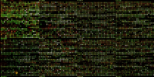
Visualised Microarray. I used this array as part of a project looking at differential methylation between head and neck cancer cells and peripheral blood mononucleocytes.

DNA that is complementary to the probes on the array will stay bound and hence show up, allowing detection. Since it is possible to put several thousands of probes for thousands of genes on an array it is easy to scan for their presence at once.

Of course, with slight modifications, it is possible to use microarrays to see which genes are being expressed in a cell. Here, RNA is isolated from the tissue and treated with Reverse Transcriptase, which produces cDNA (complementary DNA) that corresponds to the genes being expressed, cDNA is then put through the same workflow as mentioned above and one can see which genes are being expressed in the tissue sample.

Things can be taken one step further to compare variations in gene expression between different cell or tissue types or normal and diseased cells. In this case one can find out if the expression of a gene is upregulated or downregulated with respect to the reference we are using (which is called a control in scientific parlance)

Differential Expression Analysis Workflow, courtesy Memorial University, Canada.

This is done by isolating cDNA from both the control and the test sample, tagging them each with a different coloured probe, then hybridizing them on the same chip and comparing the intensities of fluorescence for each of these probes, with the composite result yielding a picture of which genes are expressed more in test sample than control and vice-versa.

This kind of procedure is also known as Array Comparative Genomic Hybridization, it can be used to assay changes in copy number too (how many copies of a gene, which translates into amounts of mRNA) to see if a gene is overamplified or deleted in a given sample.

There is an absolutely gorgeous poster on Array CGH and how it is empowering cancer research available in .PDF format from AAAS/Science here

[2] ChIP Arrays.

These are modified versions of DNA microarrays that are used to study the epigenetic status of genes, now genes can be expressed or not based on the status of the methylation of promoters and the presence of associated proteins called histones. It is possible to obtain DNA samples from tissues, isolate those that are associated with histones using an antibody that binds specifically to histones, and to then analyse these DNA samples using microarrays to find out which particular genes are subject to chromatin modification. ChIP, by the way, stands for Chromatin Immunoprecipitation.

This technique can also be used to examine the binding of other proteins such as transcription factors to DNA.

ChIP on Chip Wet lab workflow, Courtesy Wikimedia Commons.

Please click on the image for a larger version.

[3] Tissue Microarrays.

Now these kinds of microarrays are fundamentally different from the types mentioned above. These are meant solely for facilitating microscopic analysis of tissues in a manner that is more comprehensive than traditional histological methods that involve mounting one section per slide.

Tissue Microarray Slide , courtesy Wikimedia Commons.

These arrays consist of up to a thousand tissue cores in a paraffin block mounted on a slide, the idea is that examinations of multiple tissue samples can be carried out in one sweep, if you will. This is called multiplex analysis.

The process for making these is as follows.

1) Isolate tissue cores from biopsy sample using a hollow needle.

2) Embed cores in a paraffin block.

3) Section block into thin sections using a microtome.

4) Mount sections on slide.

This approach is extremely useful because tissue processing can accommodate a wide range of studies, ranging from morphological analysis to immunohistochemical studies (where the presence of certain proteins can be detected with the help of antibodies linked with stain) to fluorescence studies, where proteins can be fluorescently tagged and visualized using a fluorescent microscope.

Fluorescent Microscopy of Tissue Section, courtesy Immunoportal.

Immunohistochemically Stained Section of Bone Marrow, courtesy McGill University, Canada

Cantilever Arrays – A footnote.

Recently there have been advances in array technology that allows for the development of extremely accurate arrays which do not need the sample to be amplified or the sample to be tagged with fluorescent probes et cetera. These arrays are known as Cantilever Arrays because the probe is bound to the tip of a cantilever that can bend when complementary DNA/RNA is present in the sample. This bending results in the reflection of an incident laser beam which can be used for quantification. Now that is simple and desirable at the same time because it greatly simplifies workflows (thus saving costs) while increasing speed and reliability, which should translate to better usability in the clinic.

Cantilever Arrays and Potential Applications, courtesy Swiss Nanoscience Institute, University of Basel, Switzerland

Here is a landmark paper on the subject, titled “Multiple label-free biodetection and quantitative DNA-binding assays on a nanomechanical cantilever array” which explains what I typed above in detail. Happy Reading.

Further links and things to do

[1] You can carry out a virtual lab experiment involving differential expression analysis between cancer cells and normal cells at the University of Utah’s Genetic Science Learning Center here. Please do also peruse the additional resources they have linked to.

[2] Watch a video summarizing the basics of Microarray technology.

[3] Read a Scitable article on Genetic Diagnosis and DNA Microarrays in Cancer here

[4] Read a Scitable article on Array CGH here

I hope you have fun reading and learning.

-Ankur.

Molecular Photocopying – PCR.

Now PCR or Polymerase Chain Reaction is a vital part of many laboratory procedures involving DNA, it is a process for producing exponential numbers of copies of a given DNA molecule, that is, if you start off with one molecule of your template DNA, and run 20 cycles of amplification, you will get twenty doublings’ worth.

Before I delve into the depths of the technique, I think it is worth introducing the concept to you with the help of an amazingly funny (and appropriate) video from BioRad.

😀

Right, getting down to business now, PCR basically involves the replication of a given DNA segment in-vitro or ex-vivo(outside cells). We need a template (the DNA segment that we want to amplify) , free nucleotides needed to build new strands, a buffer containing ions to stabilize the mixture, PCR primers to help start off replication, a DNA polymerase enzyme & a method to heat and cool the reaction mixture.

The polymerase that is routinely used these days is called Taq Polymerase, this comes from a bacterium called Thermus aquaticus which lives in hot springs, this is used because it can withstand the high temperatures that are a part of the PCR process. All of the aforementioned ingredients have to be put into reaction tubes before we start cycling them through different temperatures (this is one reason that PCR machines are also called Thermocyclers). In some cases, if the Taq polymerase that is being used needs to be activated by heat, the reaction tube is heated to 96’C for about 10 minutes.

I think it is now time to move on to the process itself, which can be divided into three phases.

[1]Denaturation

In this step, the reaction tube is heated to 94-98′ C for around half a minute, this separates the double stranded DNA template into its constituent single strands.

[2] Annealing

In this step, temperature is reduced to 50′ C – 60′ C for 1 minute, this enables primers (short single stranded sequences of DNA) to bind to complementary sequences of the single stranded DNA derived from step [1] , the diagram should show this clearly. This is required because DNA polymerase requires a primer to be present in the 5′ -> 3′ direction to produce a new strand successfully.

[3]Extension

In this step, the temperature is raised to 72’C or so for 2 minutes or so, this is the optimum operating temperature for Taq Polymerase and the primers are extended such that a new double stranded copy is synthesized using the single stranded copy generated in step [1].

To sum up, we start with two strands of DNA in one molecule, which is split into two single strands which are bound to primers, followed by replication to produce two new double stranded molecules, in the next cycle we’ll start of with two double stranded molecules and end up with four, and then four and sixteen, and sixtenn and two hundred fifty six and so on, you get the idea. 😛

A video summary that explains it all is available here

Finally, there is a technique called quantitative PCR, or qPCR, which can be used to estimate how much of your given fragment is present in a sample. This works on the principle that you can have dyes that fluoresce when they bind DNA, and you can see how many cycles of amplification it takes for a certain level of fluorescence to be reached in order to figure out how much of your DNA is present in the sample. This becomes possible because you can have DNA of known concentration that permits you to work out how many cycles a particular amount of DNA will take to produce a certain amount of fluorescence, alternatively you can just compare two samples – the more template DNA you have, the quicker it’ll reach that threshold…


I’ll leave you with another funny video pertaining to PCR from BioRad.

That is it until the next post, I must say this has been one of my shorter posts of late.

– Ankur

A Little Tangent – Mantis Shrimpomania.

I am writing this intending to introduce you to the world of Mantis shrimps. They are neither Mantises nor shrimps, and the colloquial name is a misnomer, but they belong to Order Stomatopoda of the Crustaceans.

Now these animals are spectacularly aggressive hunters with some incredible cool tricks up their sleeve.They tend to stay hidden in narrow passages and burrows et cetera from which they operate as hunters that can actively chase and kill prey.

There are various biological features that make these organisms fantastic hunters, including a superb visual system. These eyes can move independent of each other, they can perceive a wide range of wavelengths, ordinary as well as polarized light. The midband, which contains six rows or so of ommatidia, has receptors of upto 16 different types, this far outstrips the range of colour perception and discrimination that is available to humans. Apparently this includes up to 12 different types of colour receptors and 4 involved in the detection of polarized light. Please see this paper for more information on some of this photoreceptorial complexity.

Note the division of the eye into three bands. The midband is especially complicated.

The really beautiful thing about each eye being divided into three units is that each eye is capable of depth perception and stereoscopic vision, which is required for visual information processing during hunting.

A lot of this processing appears to be parallel, that is, several kinds of nervous output are streamlined before being sent to the Central Nervous System, things like perception of distance boil down to an overlap in the fields of vision based on distance, which means that distance to an object can be determined by which pair of receptors in the retina the point being tracked converges on to.

A detailed treatment of visual processing in stomatopods can be found here

Of course, what use is a sniper without his rifle? Mantis shrimps are fantastic predators due to the hunting mechanisms they have evolved to use. Broadly speaking they can be grouped as smashers and spearers, based on the types of Raptorial appendages that they have (this is where the name “Mantis” comes from).

Some have sharp, spear shaped appendages, and are called spearers, and others have fat clublike appendages, and may be colloquially called smashers.

Club appendage to the left, Spear appendage to the right.

The different weapons they have at their disposal may be reflected in their choice of prey, with the former showing a preference for soft-bodied prey and the latter targeting shellfish et cetera with harder shells.

These appendages are compressed and then allowed to swing out at speed, sort of like a catapult with no projectile at the end, and it is this that does damage to prey, some of the details of Clubbed stomatopods lashing out are mind-boggling.

Compression & Energy Release Mechanisms in Peacock Mantis Shrimp , Patek et al (2004)

The aforementioned paper can be found here

The authors also write in that paper that the dactyl heel (the tip of the club) can go up to a peak speed of 23 m/s and peak acceleration of 104 km/seconds squared, that is seriously bloody quick, to the point of potentially being more than a .22 caliber bullet.

When the tip of the club is moving at a very high velocity, it builds up enormous amounts of kinetic energy, and this is what allows the raptorial appendage to do things like crack shells open. The impact force of the appendage on the target can be as much as 1500 Newtons.That can be insanely damaging.

This also has a secondary knock-on effect, resulting in compression of the water/air in front leading to the formation of bubbles that then may collapse, this effect is called cavitation. This can produce heat and light, as you will see later. If you want to read more on this you may find the apposite paper here.

To put things into perspective, the authors point out that a 2.7 mm cavitation bubble can produce 9 megapascals of pressure, which is the atmospheric pressure on venus that is apparently enough to wreck rapidly rotating boat propellers.

Now the really interesting thing about cavitation effects is that there is a shrimp, called the pistol shrimp, which snaps its claw to produce cavitation bubbles that then go on to injure and stun prey, which would be indicative of the fact that this mechanism could be damaging even when used alone. Mix in the smashing appendages of Stomatopods and you have a truly deadly combination.

I suggest you read the paper because they had these Stomatopods strike a steel surface to collect experimental data, and in my opinion that is the best feature of science…collecting actual data to find out how things work as opposed to defining ill conceived answers from thin air.

Now I want to show you something on the pistol shrimp/snapping shrimp from the good old BBC, please watch & enjoy 🙂

Now here is a demonstration of a stomatopod obliterating something and bringing its appendages into action, in beautiful detail, made possible by slow motion photography.

Mwahaha.

You can also watch a TED Talk on Dr.Patek’s work.

Baby stomatopod, awww.

I hope you have fun/had fun/will continue to have fun learning about these awesome predators.

That is all until next post, bye bye 🙂

-Ankur.