Category Archives: Stuff we’ve done.

How HPV driven cancers get their mutations…

Hi there!

It’s been a long time since I last blogged, but that is because I’ve been swimming round in data, which has incidentally led to the findings that were published in this paper , which I will describe in this post.

HPV and the link to cancer.

HPV (Human Papillomaviruses) consist of a family of viruses that infect keratinocytes (skin cells) that line the outside of the body and the inner cavities – some of them just cause warts (and genital warts) but some of them are capable of driving the formation of cancer. These types, which are called “High-risk” strains, are the ones that are targeted for prevention by HPV vaccines.

High-risk HPV strains differ from low-risk strains in terms of cancer-causing ability because of proteins they make during their life cycle. Cells need to be actively dividing to permit HPV replication and in order to do this, the virus uses two proteins, called E6 and E7 , to block and degrade two proteins in human cells, called TP53 and pRb, which are two potent tumour suppressors (genes that prevent tumour formation).

Normally, E6 and E7 are only active for a brief while during the virus’ life cycle, which culminates in the production of more viruses that restart the cycle all over again, but before HPV driven cancers form something very strange happens; by complete accident the viral genome gets inserted and integrated into human DNA in infected cells, or infected cells get locked into a state where E6 and E7 are produced all the time. Suddenly you’ve got cells with TP53 and pRb off all the time, leaving behind cells that can grow abnormally. We see this when women have cervical scrapings looked at, and see “dysplastic” cells that have grown clumpy and abnormal.

However, these dysplastic cells are not cancerous – and haven’t acquired all the hallmarks of cancer. For this to happen there need to be additional changes to the DNA sequence (Mutations) of the genes in dysplastic cells that can confer those properties. Well known examples of things that cause mutations include tobacco smoke; for quite a while it had been an open question as to where HPV-driven tumours got their mutations from.

Suspicions are aroused: could the APOBEC family of proteins be making these mutations? 

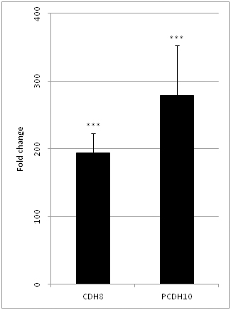
One of my major research interests is to see what genes are expressed more and what genes are turned off in HPV driven cancers, and when defining a signature for these tumours I compared them to normal tissue and HPV negative tumours that arise in the same tissue (while cervical cancers usually all tend to be HPV-driven, there are head and neck cancers caused by HPV and those caused by chronic tobacco and alcohol exposure) and one of the genes that I found expressed at high levels in HPV-positive tumours was APOBEC3B.

APOBEC3B is one of many proteins of the APOBEC cytosine deaminases family. These act either on RNA or DNA when it is a single stranded state, and take part in the body’s immune response against viruses by messing up the RNA/DNA from the viruses. They work by changing cytosines, one of the four bases that make up DNA to uracil (a base that is normally only found in RNA) which then gets converted to a thymine or a guanine (two other bases that make up DNA); so if you get lots of these changes in viral DNA you fundamentally break them so they can’t do any of the things they usually do, and it had been known for a while that you could find HPV with messed up DNA in precancerous lesions with patterns of change associated with APOBEC proteins.

This led us to wonder if APOBEC proteins could end up accidentally changing human DNA just like it would change viral DNA and therefore generate the necessary DNA sequence changes to cause cancer; and at the same time we started wondering that a couple of papers came out showing that there were human cancers in which mutations looked like they were being generated by APOBEC enzymes, very likely APOBEC3B (We could tell it was likely APOBEC 3B because it is known to change cytosines that are preceded by a thymine and followed by guanine or adenine or thymine, so if the sequence was TCA or TCG or TCT it would be converted to TGA/TTA or TGG/TTG or TTT/TGT ). There is an alternative process that can also generate TCG->TGG/TTG mutations, so in order to specifically measure APOBEC activity we ended up using the others, which we referred to in the paper as TCW to TKW (TCW->TKW, where K = G or T and W = A or T).

Those previous papers also noted that cervical cancers had lots of mutations that showed the APOBEC signature, but the question remained – was this down to it being the cervix? or was it down to these tumours being HPV+? We decided to take a look in head and neck cancers as well where we could compare HPV+ and HPV- tumours that arose in similar tissues to see if there was truly an association with HPV, and hence we did the work reported in the paper…

HPV positive tumours have a vastly higher fraction of mutations belonging to the APOBEC signature.

First, we ended up looking at levels of APOBEC mutagenesis and how much of all the mutations in tumours were attributable to them using publicly available data for 40 HPV+ head and neck tumours and 253 HPV- head and neck tumours. To do this we used multiple approaches – including looking at TCW->TKW mutations and also trying to break down all the mutations we see in these tumours into patterns of mutations, as was done by these people at the Sanger Institute , and also looking at enrichment for the TCW->TKW mutation pattern locally. All the approaches we used showed the same thing – HPV+ tumours had a vastly higher proportion of mutations most likely caused by APOBEC enzymes.

Figure1:APOBEC mutations are highly enriched in HPV+ HNSCs

Multiple measures of APOBEC activity showed a strong association with HPV status but not age or smoking; APOBEC, age and smoking were the three processes we identified as driving the signatures using the Sanger Institute’s approach. The more the numbers are shifted to the right the stronger the association with the factor listed on the left. 

We found signatures previously associated with APOBEC, smoking and age, and showed that APOBEC activity was not associated with the latter two, which was as expected. Having identified an association with HPV driven tumours we wanted to know if this was a general antiviral response or something HPV specific…so we took a look at patterns of mutations in liver cancers caused by hepatitis B and C viruses and found no evidence for APOBEC mediated mutations being significantly enriched in these tumours.

Of drivers and passengers

Most tumours have hundreds and thousands of mutation, but only a few actively contribute to the acquisition and maintenance of the hallmarks of cancer. So, having initially identified high proportions of APOBEC-mediated mutations in HPV driven cancers when looking across the exome (all protein coding genes in general) we decided to ask if the enrichment we saw in all genes was also maintained when we restricted our searching to genes known previously to drive cancer or those that share features associated with drivers, like occurring at a frequency greater than expected by chance. Our analyses confirmed that APOBEC-mediated mutations were again enriched in the HPV+ head and neck, and cervical cancers compared to the HPV- HNSCs.

Figure2

Differences between HPV negative HNSCC and HPV+ tumours (HNSCC and Cervical cancer) are maintained when looking at all protein-coding genes (whole exome) and likely driver mutations (MutSig).

Then we went on to look at which driver genes happened to be most mutated by APOBEC proteins, and found a gene called PIK3CA (one of the components of a protein complex called PI3 kinase) towards the very top of the list. PIK3CA has previously been reported as being vital to the sustenance of many HPV positive tumours in particular and head and neck cancers in general, and drugs are being developed to target it. Interestingly, we observed that in the HPV+ tumours 22/25 PIK3CA mutations recorded were of the APOBEC type, while this wasn’t the case for the HPV negative tumours.

This then led to yet another question – can the levels of APOBEC activity explain a preference for APOBEC mutations in HPV-positive tumours? Now for driver genes there are two things that may govern what kinds of mutations we see – how much of a growth advantage a mutation in a driver gene gives that cell and the mutation itself. My supervisor, Tim Fenton, who worked on PI3 kinases previously, knew that there were two regions in PI3 kinase amongst which mutations regularly occurred (one or the other) and then realised that one of them contained a TCW sequence that APOBEC proteins could act on while the other one did not.

The PIK3CA gene makes a protein called p110-alpha, and proteins have different distinct elements in their structure, called domains. One region, called the helical domain, is often mutated at two TCW sequences while the other region, called a kinase domain, is not, and both mutations confer similar growth advantage, and if you look across multiple tumour types, overall you tend to see a 50-50 split between the two. This enabled us to account for growth advantage and directly see if APOBEC activity, which we had already measured by looking at all protein-coding genes, and a preference for APOBEC-induced mutations in the helical domain, were linked.

Since PIK3CA is mutated in multiple types of cancers, I was able to grab some data from The Cancer Genome Atlas project and measure how strongly there was a skew towards acquiring helical domain mutations compared to the kinase domain mutations and just look at what APOBEC activity looked like in each of those types of tumours. The results were quite robust – the higher the APOBEC activity in a cancer type, the stronger the preference for helical domain mutations compared to kinase domain mutations.

Figure3

Figure 3. A – as you move from left to right (tumour types are arranged from left to right based on median APOBEC activity), you see helical domain mutations (black bars) become strongly preferred compared to kinase domain mutations (yellow bars). B – plotting the median TCW->TKW fraction (APOBEC activity) against the proportion of PIK3CA mutations that are helical hotspot mutations shows a strong correlation.

So yeah, people had been wondering why in bladder cancers, for example, you saw such a strong preference for helical hotspot mutations – we basically addressed that long-standing question with these analyses.

Explanatory factors

So the one other thing we did was to look at what might be driving this process, and surprisingly we found no correlation between how much E6 and E7 was being expressed in these tumours and APOBEC activity, or for that matter between APOBEC3B gene expression and APOBEC activity, and did find a strong link with how many mutations in total these tumours had. The work has led us to hypothesize it may be something like DNA damage induced by HPV, that generates the substrate for APOBEC3B to act upon, that drives the process.

Conclusion

Our work suggests that HPV positive tumours evolve in a trajectory where they incorporate HPV DNA into their own, leading to sustained E6/E7 expression, followed by APOBEC activity until a driver mutation occurs, after which clones expand and show the APOBEC signature when their DNA is sequenced while in HPV negative HNSCC smoking and alcohol do this job, and if PIK3CA is the gene mutated the HPV positive tumours tend to have helical domain hotspot mutations because APOBEC proteins are responsible for them…

Additional stuff

The journal did a Q&A that expands on some of the work in the paper, and you may find it here .

There is a press release from UCL here.


Paper Review: Identification and functional validation of HPV-mediated hypermethylation in head and neck squamous cell carcinoma

Hello everyone.

It has been bloody long since I last blogged because I have been battling illness and getting stuck into research at the same time. I’m going to review a paper I did some work towards in this post. I have written about DNA methylation in the past and the research groups I worked with/continue to work with were focusing on the various genetic and epigenetic alterations that characterise  head and neck squamous cell carcinoma (HNSCC), which is the sixth most common type of cancer.

HNSCC can be thought of as two distinct cancers with vastly different prognosis and aetiologies; a vast majority are caused by heavy smoking overlapping with heavy drinking and an increasing proportion is caused by HPV infection, transmissible through oral sex. (HPV, by the way, is the same virus that drives cervical cancer). HPV infection per se is insufficient to cause the cancers associated with it – there have to be additional genetic and epigenetic modifications on top. HPV positive HNSCC has excellent survival relative to HPV negative HNSCC, by the way.

In this study, we obtained clinical samples of both HPV positive and HPV negative HNSCC – some were fresh frozen upon surgical resection/biopsy, a lot were FFPE samples (Formalin fixed,paraffin embedded) and we also profiled cell lines using Illumina 450k methylation arrays, which give a read-out of methylation at 483,000 CpG sites (A cytosine followed by a guanine) across the human genome for less than a large pizza per sample.

The FFPE samples were used as a training set and the Fresh frozen samples and cell lines were used as a validation set. We found quite a few interesting things…

[1] HPV positive HNSCC exhibits a greater degree of DNA methylation (Hypermethylation) than HPV negative HNSCC, especially in genes that are known to be silenced by PRC2 complexes in stem cells. PRC2 complexes consist of multiple proteins that co-operate to produce the H3k27 histone mark. We find the same genes being silenced by DNA methylation instead in HPV positive HNSCC. This is also strongly associated with differences in expression; More the methylation, less the expression, as it should be.

HPV positive HNSCC exhibits hypermethylation relative to HPV negative HNSCC. Blue represents high methylation and yellow represents low methylation.

HPV positive HNSCC exhibits hypermethylation relative to HPV negative HNSCC. Blue represents high methylation and yellow represents low methylation.

[2] A subset of HPV positive HNSCC showed very high degrees of methylation – which is called a CpG Island Methylator Phenotype, and was associated with significantly worse survival.

CIMP phenotype (Cluster 1a) is associated with very high methylation, HPV positivity and significantly worse survival compared to HPV positive tumours with comparatively less methylation (Cluster 1b) as shown in the Kaplan Meier curve at the right.

CIMP phenotype (Cluster 1a) is associated with very high methylation, HPV positivity and significantly worse survival compared to HPV positive tumours with comparatively less methylation (Cluster 1b) as shown in the Kaplan Meier curve at the right.

[3] If you put the viral oncogenes E6 and E7 into a cell line that was derived from HPV negative HNSCC, you tend to see that E6 induces hypermethylation. This wouldn’t be surprising because p53, which is blocked by E6, is known to regulate DNMT1, a DNA methyltransferase that is involved in the maintenance of methylation.

[4] If you use probes on the array that are significantly different between HPV positive HNSCC and HPV negative HNSCC, and compare them to publicly available data for cervical and lung cancer by a process called multidimensional scaling, you find that HPV negative HNSCC is closely related to lung cancer while HPV positive HNSCC is closer to cervical cancer, suggesting that HPV modulates the methylation patterns that make cervical cancer closer to HNSCC of this type.

Multidimensional scaling shows HPV negative HNSCC (HPV0) to be more closely related to lung cancer and HPV positive HNSCC to be similar to cervical cancer.

Multidimensional scaling shows HPV negative HNSCC (HPV0) to be more closely related to lung cancer and HPV positive HNSCC to be similar to cervical cancer.

[5] The relationship between methylation and expression is valid and as predicted even in a panel of HNSCC cell lines, as I demonstrated using qPCR, where we get RNA, make DNA, and then do PCR to find out how many cycles it takes to get past a particular threshold of fluorescence.

Genes that are significantly hypermethylated in HPV positive HNSCC are relatively overexpressed in HPV negative HNSCC as expected (The relationship between most methylation and expression is inverse)

Genes that are significantly hypermethylated in HPV positive HNSCC are relatively overexpressed in HPV negative HNSCC as expected (The relationship between most methylation and expression is inverse)

[6] We found that DNMT1 and DNMT3a , which are enzymes involved in maintaining and establishing DNA methylation, are expressed more in HPV positive HNSCC cell lines relative to HPV negative HNSCC cell lines as a group.

DNMT1 and DNMT3a are significantly overexpressed in a panel of HPV positive HNSCC cell lines vs HPV negative HNSCC cell lines.

DNMT1 and DNMT3a are significantly overexpressed in a panel of HPV positive HNSCC cell lines vs HPV negative HNSCC cell lines.

So basically, we started off with two subsets of a type of cancer, identified that the methylation patterns between them are different, that this has functional ramifications and clinical implications. It would be very interesting if someone ended up looking at hitting methylation in HPV positive cancers with anti-methylation drugs to see if that high level of methylation is just an artefact of how HPV positive HNSCC develops or whether there are therapeutic opportunities to be had.

Journal Reference 
Matthias Lechner, Tim Fenton, James West, Gareth Wilson, Andrew Feber, Stephen Henderson, Christina Thirlwell,Harpreet K Dibra, Amrita Jay, Lee Butcher, Ankur R Chakravarthy, Fiona Gratrix, Nirali Patel, Francis Vaz, Paul O’Flynn, Nicholas Kalavrezos, Andrew E Teschendorff, Chris Boshoff and Stephan Beck, Identification and functional validation of HPV-mediated hypermethylation in head and neck squamous cell carcinoma, Genome Medicine 2013, 5:15 doi:10.1186/gm419
URL – http://genomemedicine.com/content/5/2/15

Cheers,
Exploreable.

Examining the cytotoxicity of metallic nanoparticles…a little blast from the past.

This is just a little experiment I did with a classmate during my undergraduate years to see if metallic nanoparticles could have an effect on bacteria and yeast, which could perhaps be used to address the use of nanoparticles in a microbicidal capacity.

Here goes the report.

Abstract

Introduction

An investigation was carried out on the cytotoxic properties of metallic nanoparticles on both Prokaryotic and Eukaryotic model organisms using qualitative assays for cytotoxicity.

Methodology & Results.

Metallic nanoparticles of Copper, Silver and Iron were prepared using Turkevich synthesis, these were then screened for cytotoxicity using well diffusion assays using a reference set of four bacterial species, results indicated that Silver nanoparticles were potently cytotoxic against bacteria, being capable of inducing substantial zones of inhibition in the medium into which said particles could
diffuse.

The test was repeated for Eukaryotes using Saccharomyces cerevisiae as the model organism and turbidimetry for evaluation. Values were obtained as a function of varying concentration vs varying turbidity. Results indicated a direct statistical relationship between the strength of nanoparticle
solutions of Silver as opposed to the other two.

Conclusions
The cytotoxicity of nanoparticles of silver was demonstrated in both prokaryotes and eukaryotes, this backed up the previously reported instances of nanosilver toxicity in the scientific literature & highlighted the potential concerns associated with environmental contamination of the environment
with metallic nanoparticles. It also raised questions about the potential use of silver nanoparticles in conjugates as a form of cytotoxic therapy, which may be worth investigating.

Materials and Methods

Turkevich Synthesis

Materials Required

0.1 M Silver Nitrate solution, 0.1 M Copper Sulphate Solution, 0.1 M Ferrous sulphate solution, 38.8 M Sodium Citrate solution (aqueous) , deionized water, standard glassware.

Principle

Turkevich synthesis uses a reducing agent to first reduce metal salts to nanoparticles, the same agent then inhibits re-coagulation.

Procedure

Solutions of the aforementioned nature were prepared from raw salts using deionized water, then reaction mixtures were prepared using metal salt solutions and sodium citrate in the ratio 2:1.

A water bath was heated to 100’C and the reaction mixtures were boiled until colour changed , this change in colour is often attributed to the Surface Plasmon Effect, which is due to changes in the dielectric properties of synthesized nanoparticles.

The solutions, once colour had changed, were gradually cooled back to room temperature and stored in reagent bottles, estimation of the concentrations of the nanoparticles and shape-size characterization weren’t carried out due to the lack of access to a Scanning Electron Microscope.

The production of nanoparticles was verified by cross-corroborated change in colour due to the surface plasmon effect.

Assay for Prokaryotic Cytotoxicity

Materials Required

Bacterial broth cultures, 24-48 hours old , of Staphylococcus aureus, Bacillus subtilis, Enterobacter aerogenes and Escherichia coli, cork borer, nutrient agar/blood agar base medium, laminar airflow unit, swabs, micropipettes and pipette tips, Ethanol (70%), standard glass labware.

Procedure

All non-heat labile materials were sterilized by autoclaving under standard conditions, 121’C at 15psi, 20 minutes, heat labile materials such as pipettes were sterilized using ultraviolet irradiation. Sterile, aseptic conditions were established for carrying out the experiment

Swab cultures were carried out and a well diffusion assay was set-up using 75 microliters of the nanoparticle dispersions in the first round and a much higher concentration of 200 microliters in the second phase of the experiment, in the preliminary, first round, just E.coli and S.aureus were used, and the well diffusion assay took into account dilutions of nanoparticles (x, x/10 & x/100) , it was found that preliminary antimicrobial activity of any significant potency was only shown by the dispersions at their original concentrations, therefore this was the only concentration chosen for further corroborative investigations.

Results of prokaryotic cytotoxicity assay. Zones of inhibition are clearly apparent.

The process was repeated the second time with all four of the aforementioned cultures, with only original concentrations of the nanoparticle dispersions being used.

Nansosilver at the original concentration showed the maximum amount of microbicidal activity, which by inference makes it cytotoxic to prokaryotes, data about ZOI diameters is available in the appendix that follows this report.

Assay for Eukaryotic Cytotoxicity

While the initial plans included the use of trypan blue cell exclusion method for corroboration, the inconsistency of the yeast dispersion used meant that the results were practically unusable, and therefore data had to be tentatively evaluated using turbidimetry alone, we recommend caution before jumping to conclusions on the basis of this experiment alone and suggest further corroboration and verification.

Materials

yeast dispersion, 1 pellet in 50 ml of sucrose broth (approximately 0.5 g added) , nanoparticle dispersions with dilutions (x , x/10 and x/100) respectively, photocolorimeter (a spectrophotometer was preferable but not available)

Procedure.

Control and test solutions were created for each set of nanoparticle dispersions, for all three metallic nanoparticles, the controls consisted of 1 ml of the nanoparticle dispersions, and 1 ml of uninoculated sucrose broth and 8 ml distilled water, while the test samples contained 8 ml distilled water, 1 ml nanoparticle solutions and 1 ml of inoculated sucrose broth, the tubes were plugged with cotton and incubated overnight at room temperature (25-27’C)

Readings for optical density were then taken after 24 hours of culture, to examine if there was a trend between dilutions and rate of cell growth, a direct relationship between the two variables would yield an inversely proportional relationship.

A trend of cytotoxicity was found to occur for treatment with nanosilver solutions, this has been corroborated by studies which have illustrated the ability of silver nanoparticles to induce mutations and cell death (P V Asharani et al 2008 Nanotechnology) The other two nanoparticle dispersions showed no cytotoxicity.

The raw data and the graphical analysis of the data from this experiment is also available in the appendix at the end, a baseline correction factor was included to make presenting information in the graphs easier, the correction was applied by adding an equal amount to all derived results.

Results and Conclusions

Turkevich synthesis can be carried out in simple facilities to produce nanoparticle dispersions for further investigation.

Silver nanoparticles are potently cytotoxic against all the organisms tested against in the prokaryote cytotoxicity assay, regardless of whether they are gram +ve or gram –ve, and as such can be used as microbicides.

However, silver nanoparticles have been shown to be cytotoxic against Eukaryotes too, this means that unregulated efflux into the environment can lead to disastrous ecological consequences, and therefore strict regulatory norms are imperative.This cytotoxicity, however could have biomedical applications in the form of immunoconjugates against cancer specific receptors, for instance,

The other nanoparticle dispersions (Copper and Iron) , at the tested concentrations, were found not to be cytotoxic , as were silver dispersions at less than x/10 dilution, this could indicate a preliminary statistic for hazard levels and an association with the concentration, while clearing the other two for free usage at the investigated concentrations.

However, for the data to become fully valid for policymaking, an accurate method such as mass spectrometry must be used to evaluate precisely the concentration of the nanoparticles in their respective dispersions.

To sum up, our data is in agreement with research findings that silver is cytotoxic, and these studies so far point towards iron and copper not being cytotoxic, however, further research needs to be carried to account for any imperfections in the study itself; these results are a rough starting point, at best.

References

P.K. Khanna, Narendra Singh, Deepti Kulkarni, S. Deshmukh, Shobhit Charan, P.V. Adhyapak, Water based simple synthesis of re-dispersible silver nano-particles.

Iron nanoparticles: Synthesis and Applications in surface enhanced Raman Scattering and electrocatalysis, Guo et al, Physical Chemistry Chemical Physics, 2001.

An antibiotic assay by agar well diffusion method, Perez et al, Acta Biol. Med. Exp, 1990

Trypan Blue Exclusion Test of Cell Viability , Strober, Current Protocols in Immunology,

Toxicity of silver nanoparticles in zebrafish models
P V Asharani et al 2008 Nanotechnology 19 255102 (8pp) doi: 10.1088/0957-4484/19/25/255102

Appendix A : Turbidimetry Data and Graphs

Results from turbidimetry experiment.

Appendix B : Prokaryotic Cytotoxicity Assay.

Prokaryote zone of inhibition data.

Appendix C: Tentative estimates of concentration.

Silver Nanoparticle dispersion.

100 ml of AgNO3 solution contained 10.8 mg of Silver
3 ml of the finally prepared solution contained 0.324 mg of Silver nanoparticles

Solutions for the Yeast assay, contained 0.108 mg (x) , 0.0108 mg (x/10) and 0.00108 mg (x/100) of
Silver & 200 Microlitre aliquots, used for the prokaryote assay, contained 0.0216 mg of silver

Iron solution.

100 ml of FeSO4 solution contained 5.6 mg of Iron

3 ml of the prepared nanosolution contained 0.168 mg of Iron Nanoparticles
1 ml, for the yeast assay, contained 0.056 mg (x) , 0.0056 mg (x/10) , 0.00056 (x/100)

200 Microlitre aliquots, for prokaryote assays, contained 0.0112 mg of Iron nanoparticles


Copper solution.

100 ml of CuSO4 solution contained 6.35 mg

1 ml of the prepared solution contained 0.0635 mg (yeast assay) , the other two quantities used were 0.00635 mg and 0.000635 mg.

200 Microlitre aliquots contained 0.0127 mg